Book Review: Vertebrate Skeletal Histology and Paleohistology
Reviewed by James Farlow (Purdue University Fort Wayne, Fort Wayne, IN)

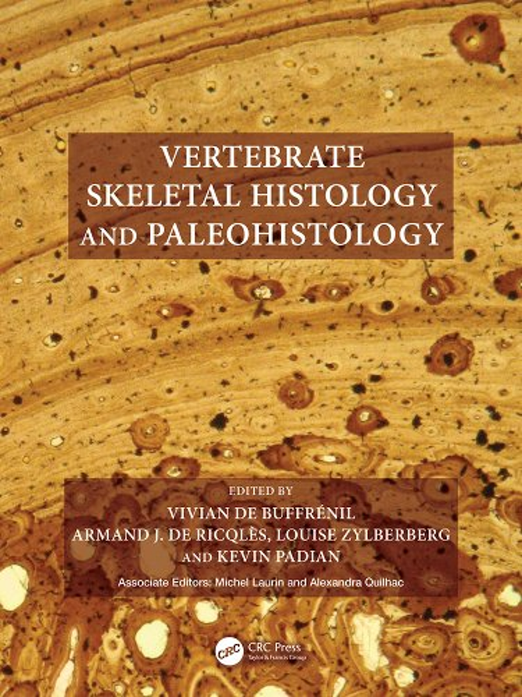
de Buffrénil, V. A. J. de Ricqlès, L. Zylberberg, and K. Padian (Editors), M. Laurin and A. Quilhac (Associate Editors). 2021. Vertebrate Skeletal Histology and Paleohistology. CRC Press, Boca Raton, Florida, 825 pp.
This is a BIG book: 825 pages long, weighing in at 5 pounds. Collectively there are 40 chapters, written by 45 specialists (a gallery of the most authoritative researchers in the field of vertebrate skeletal histology). Each chapter has its own extensive bibliography.
I read this book as someone who knew a bit about, and had dabbled a bit in, vertebrate paleohistology without considering myself an expert. I wanted to learn more about this area of research, the current state of the art. This book provides all that and more; as its title suggests, there is useful information here not only for paleontologists, but also for specialists on the osteohistology of extant vertebrates, and possibly even medical personnel. Much of the material in the book is quite technical, and so novice readers (like me) will likely have to work a bit to digest it.
The book is organized into six sections. A short first section (Chapter 1) about the history of the field is followed by five sections that provide the meat of the book.
The second section is a general survey of vertebrate skeletal morphology and histology. I think this will be the most useful section of the book for newcomers to the field of bone histology, who will likely frequently refer back to this section while reading chapters later in the book. I also think this section will be very useful to persons teaching introductory courses in comparative anatomy and vertebrate paleontology. Among the topics considered are: the embryonic development of the skeleton; a very nice survey of different regions of the vertebrate skeleton; discussions of microanatomical features and tissue types of bone; descriptions of cartilage histology; the relationship between bone cells and the organic matrix that deposits them; processes of mineralization of bone; and the relationship between bone tissue type and accretion rates. Interspersed among the descriptive material are short sections about the current research methodology used for studying the features of those chapters.
The texts of this section are very good, of course, but the illustrations are outstanding. I was particularly taken by Figures 2.1 (diagrams of embryonic development of skeletal tissues); 2.4 (diagram and photographs of tetrapod limb development), all of the figures in Chapter 3 (diagrams and photographs of the regions of the vertebrate skeleton); 4.8 (diagram of vascular network patterns in bone cortex—I referred to this figure many times as I worked my way through the book); 8.1 (photographs of cross sections showing contrasts between bone cortex and medulla, periosteum and endosteum, and primary vs. secondary bone); 8.4 (thin sections of bone cortices showing vascular network patterns); and 8.8 and 8.11 (thin sections of different bone tissue types).
There is so much information presented in the remaining sections that a proper summary of their contents would be a book in itself. Consequently, I will offer only brief summaries of things that stood out to me.
Section three deals with processes of bone formation, increase in size and shape during bone growth, accretion rates, processes of remodeling, and formation of metaplastic tissues like osteoderms and calcified tendons. Section four is a single chapter dealing with hard tissues of teeth of mammals. Dentine, enamel, and cementum are described and extensively illustrated with diagrams, photographs, and SEM images. There are discussions of periodic growth lines in dental tissues, as well as the authors’ lengthy evaluation of preparation and imaging techniques that they like or dislike. I think that this chapter will be useful to wildlife biologists as well as paleontologists.
Section five constitutes about a third of the book, and surveys the skeletal tissues of the various vertebrate clades. It begins with a very short introduction (Chapter 14) that nevertheless provides two useful diagrams, the first of which (Fig. 14.1) shows the phylogeny of all vertebrates, but with emphasis on fishes (finned vertebrates; also see Figs. 15.1 and 15.2), and the second (Fig. 14.2) of tetrapods (stegocephalians). This is followed by 15 chapters that work their way “up” vertebrate phylogeny: finned vertebrates, basal tetrapodomorphs, lissamphibians, basal amniotes, testudines, lepidosaurs, marine reptiles, archosaurs and their close kin, nonmammalian synapsids, and mammals. Each chapter begins with a useful summary of the currently understood phylogeny of the clades discussed in that chapter, followed by a survey of the histology of hard bone tissues in that group.
For many groups the distribution of bone tissue type, vascularity, and skeletal growth marks is used to infer absolute or relative bone accretion rates and overall animal growth rates and, by inference, the metabolic rates necessary to support those rates. Faster growth rates than expected for most extant reptiles are inferred for pareiasaurs (p. 379), leatherback sea turtles (p. 387), large varanids (pp. 401–402), mosasaurs (p. 416), some placodonts (p. 430) and pachypleurosaurs (pp. 437, 441), non-archosaurian archosauromorphs (pp. 473, 476–477), and most non-mammalian synapsids (p. 560), with even faster accretion rates, implying tachymetabolic endothermy or something close to it, in plesiosaurs (pp. 453–455), ichthyosaurs (p. 464), some non-archosaurian archosauriforms (pp. 476–477), some non-ornithodiran archosaurs (pp. 477, 482), ornithodirans (pp. 482, 542), and of course mammals and their closest relatives. Interestingly, crocodylomorphs (including extant crocodylians) have a bone histology that suggests they have reverted from the higher resting metabolic rates of their close Triassic relatives to a thermometabolism more like that of other living reptiles (p. 505).
Section 6 comprises 11 chapters that focus on “integrative questions” of biomechanics, ecology, and thermophysiology that concern more than one vertebrate clade: whether patterns of osteohistology shed light on phylogeny; the utility of skeletal growth marks for determining the age of the animal, and whether it was still growing, at the time of death; responses of bone to mechanical stress; correlation of bone (stylopodial, ribs, vertebrae) microanatomy (relative thickness/compactness of cortex vs. medulla/spongiosa) with lifestyle (terrestrial, arboreal, fossorial, volant, amphibious, or aquatic; correlation between bone histology and resting metabolic rate; functional significance of bone ornamentation (e.g. osteoderms); and application of bone and tooth histology to paleoanthropological and archaeological research questions. I was particularly interested in reading the chapter entirely devoted to the relationship between microanatomy and lifestyle, which documents remarkable convergence in bone microanatomy and vascularity across several clades of secondarily marine forms.
For me, the greatest value of the book comes from the sheer number of photographs and other images of thin sections of different bone tissues from different interior regions of limb bones. The cumulative impact of images helps considerably in getting an idea of what a particular bone cortical texture and degree of vascularity looks like.
This book is so impressive and useful that I am hesitant to offer any criticism, but I wish it had included an index. The editors append an extended table of contents at the end of the book “so that readers can easily identify the types of tissues that interest them and, by reading those sections, the types of animals in which they appear as well as the overarching questions of the field” (p. 799). Fair enough, but this book is so big that it took me several months to work my way through it. In some chapters I often found myself trying to remember what a particular cortical tissue type, or pattern of vascularity, was like, or the meaning of a histological term or acronym (e.g., MAR: mineral accretion rate; BFR: bone formation rate; CCCB, compacted coarse cancellous bone), and having to flip through several pages before I found the description or figure that would jog my memory. It would have been nice to have an index that would tell me instantly what page(s) dealt with the topic I was looking for. In addition, or alternatively, a glossary of terms and acronyms, like Table 4.1’s treatment of bone microanatomical features, but for the book as a whole, would have helped.
A minor criticism is that the binding of the book (at least in the review copy I received) isn’t strong enough to support the book’s size and weight. It didn’t take long for the hard cover to come loose over the book’s spine, and for the spine itself to crack.

